Contect
- From your Kitchen to Clinical Settings.
Introduction
Did you know that microwaves are not only used to heat your food? In the biomedical industry this technology can measure tissue changes, guide minimally invasive treatments and even monitor vascular implants such as stents without ionizing radiation. In recent years, the use of microwaves has been gaining traction in biomedical research and the healthcare sector because it combines three attributes that are hard to bring together: portability, safety, and strong matter interaction capacity.1
What are they?
Microwaves are electromagnetic waves that occupy the frequency range between 300 MHz and 300 GHz, lying in the spectrum between radio waves and infrared radiation.2
Also, microwaves are a type of non‑ionizing radiation. Unlike X‑rays, they do not have enough energy to break chemical bonds or directly damage cellular DNA, which makes them intrinsically safer for diagnostic uses.3,4 This non-ionizing nature is specifically valuable for repeated follow-up applications such as monitoring patients with implanted devices.
Before medical applications, microwaves transformed industrial processes through volumetric heating. From food processing, providing rapid tempering of frozen blocks,5 to advanced materials synthesis like technical ceramics6,7 and green chemistry, nanomaterials, and pharmaceutical synthesis,8,9 microwaves reduced energy and production times.
Medical applications
New Applications and Clinical Perspectives
In healthcare, microwaves are increasingly explored for two main directions: (i) therapeutics, using controlled energy delivery to heat targeted tissue (e.g., ablation or hyperthermia, and (ii) imaging and sensing, using low-power measurements to detect dielectric or electromagnetic changes.
After decades of research into medical microwave applications, the last few years have brought a significant shift: from conceptual validations toward real clinical devices that are beginning to transform medical practice.
Therapeutics: Ablation, Hyperthermia and Surgical Monitoring
Therapeutic microwave applications are the localized, controlled transmission of electromagnetic energy to raise the temperature of the target tissue. Two clinical approaches dominate the field: hyperthermia and thermal ablation.1,10 Ablation uses high temperatures (typically ≥60 °C applied for brief intervals) to induce irreversible tissue necrosis and is an established strategy for treating hepatic, renal, bone, and other tumours. On the other hand, hyperthermia uses moderate temperatures (39–45 °C) to sensitize tumour tissue to concomitant radiotherapy, enhancing cytotoxic effects via mechanisms such as inhibition of DNA repair and sensitization of hypoxic tumour cells.11
Microwave‑based technologies have been proposed as one of the most versatile platforms for controlled induction of deep hyperthermia, thanks to their ability to deposit energy locally and adaptively.10 The clinical appeal is clear: localized control without incision, potential for outpatient procedures, and, when combined with imaging, the ability to guide therapy in real time. However, as with any thermal therapy, the main challenge is ensuring sufficient dose without collateral damage to adjacent structures.
Diagnosis: Microwave Imaging and Sensing
Fundamental Principles
Using the dielectric contrast between tissues, microwave imaging aims to reconstruct internal information from the human body. Dielectric contrast arises because changes in water content and tissue structure fundamentally modify how electromagnetic waves propagate, scatter, and reflect.
The field of microwave imaging has undergone rapid development over the past five years, with recent reviews documenting the state of the art in antenna configurations, reconstruction algorithms, and clinical validation.1 These technologies do not aim to replace other diagnosis methods like mammography or MRI in all contexts, but rather to offer specific advantages where those traditional modalities face limitations.
Monitoring and Sensing: From Diagnosis to Follow‑up
Near‑field Sensing vs Imaging
Although both use microwave signals, imaging and sensing differ in what they output and how they are used clinically: (i) Microwave imaging aims to reconstruct a spatial map (an image) of dielectric properties inside the body. Its goal is localization and characterization (e.g., “where is the lesion and what does it look like?”). This typically requires many measurements, complex reconstruction, and careful control of geometry.
Microwave-based clinical imaging devices show promising results, with high performance in breast cancer detection,12–16 stroke detection systems, identifying small haemorrhages and already in clinical trials1,10 and new applications for in kidney screening, highlighting portability and safety.17
(ii) Microwave sensing aims to detect a signature or change in electromagnetic response associated with a clinical stage (e.g., “is something changing over time?”). It may not produce an image at all; instead, it produced quantitative readout or classification that supports monitoring and decision making. In practice, imaging is often used for one-time diagnostic assessment, while sensing is well suited for longitudinal follow-up, where safe repeatability and consistency across visits matter as much as spatial resolution.
A particularly relevant line of work is the use of microwaves for near‑field sensing. This approach does not aim to generate a CT‑like clinical image, but to detect measurable changes in signals scattered by the body or by an implanted device.1 The conceptual shift is important. Instead of reconstructing the full anatomical structure, the aim is to detect electromagnetic changes that correlate with specific clinical events.
This approach has been reviewed recently for applications such as stroke diagnosis and body implants (stents), cancer screening, and continuous monitoring of biological parameters.1,10,18 The main challenge is typically not limited to hardware (antennas, skin coupling, transmitted power), but to software (robust algorithms, adaptive calibration, and AI integration).1 Emerging sensing applications include intracranial pressure monitoring, cardiac and bladder sensors, smart contact lenses, and wireless multichannel neural interfaces.10 While hardware constraints remain relevant, current challenges increasingly involve software, including robust algorithms, adaptive calibration, and AI integration.1
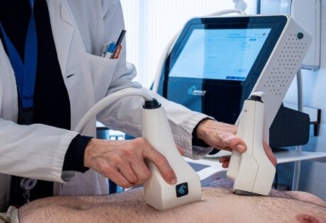
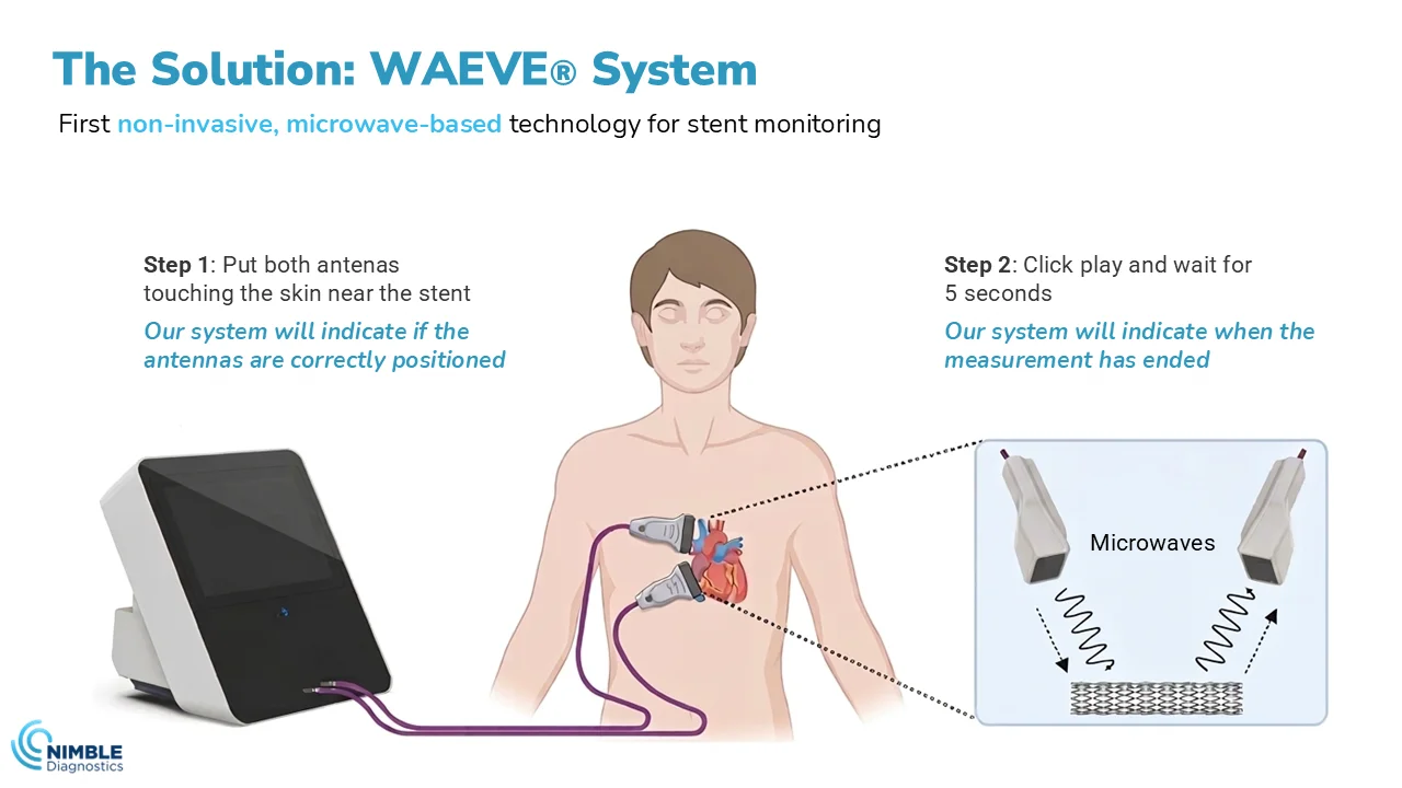
NIMBLE Diagnostics represents a practical application of microwave sensing for cardiovascular implant monitoring. Preclinical validation demonstrated that microwave interferometry detects stent resonance shifts correlating with structural changes (recoil, fracture) and biological deposits (cholesterol, thrombus) in animal models and phantoms. Building on this foundation, the technology has progressed into early clinical studies, marking an important step toward clinical adoption.
By enabling simple, non-invasive, repeatable follow-up of millions of stent patients worldwide, NIMBLE could reduce reliance on invasive procedures, improve early detection of complications, enhance patient safety, and generate substantial savings for health systems.
Advantages of Microwave‑based Technologies
Microwaves offer significant advantages over traditional diagnostic technologies. Unlike X‑rays and CT, they do not involve ionizing radiation, removing safety barriers for repeated screening, especially in paediatric and pregnant populations.1,10 In longitudinal follow‑up contexts such as stent monitoring, this advantage compounds. While CT angiography limits the number of safe procedures due to cumulative risk, microwaves can be used indefinitely without radiological concern.1,3
Microwave systems (with antennas + RF electronics) can be miniaturized more easily than CT or MRI systems, enabling point‑of‑care diagnosis and deployment outside the hospital.1,10
In therapeutic applications, microwaves allow localized, controllable heating, depositing focused energy with thermally optimized profiles that can be computed and validated in real time.1,10 Current research lines focus on optimizing field distribution and temperature control for precise hyperthermia.19,20
Finally, the signal nature of microwaves, with scattering and impedance parameters, naturally lends itself to advanced algorithmic analysis. Machine learning has shown superiority over deterministic reconstruction in classifying pathologies and compensating for anatomical variability, paving the way for next‑generation hybrid MW‑AI systems.21
Real‑world Challenges and Limitations
Although the potential of medical microwave technologies is considerable, they face significant technical limitations.1 Most systems operate between 1–15 GHz and depend critically on wave–tissue coupling, which varies with frequency, body geometry, skin contact quality and individual anatomical variability, requiring meticulous calibration and, preferably, personalized baselines for longitudinal follow‑up. Furthermore, in portable devices, achieving reproducible contact between skin and sensor is a major technical challenge.1
Image reconstruction is an intrinsically challenging inverse problem where multiple dielectric distributions can explain the same measurement, making the solution particularly sensitive to noise and artifacts.1,22 Machine learning has shown an ability to reduce reliance on explicit physical modelling, but introduces serious regulatory challenges due to algorithmic opacity in clinical devices.1,21 In sensing, a practical strategy is to focus on patient-specific baseline and robust change detection over time.
Interindividual biological variability aggravates these complications: dielectric properties vary by ±25–40% depending on age, hydration, body composition, and local pathology, making population reference databases inconsistent.1 Although individualized calibration technically addresses this issue, it significantly increases operational complexity.
The fundamental trade‑off between spatial resolution and penetration further limits practical applications: high frequencies achieve ~mm resolution but penetrate barely 1 cm, while low frequencies reach ~10 cm but sacrifice resolution to 2–5 cm. This compromise explains why microwave imaging has not replaced mammography in dense breast tissue, where deep penetration and precise localization are simultaneously needed.1
All these technical challenges are compounded by strict regulatory burdens requiring robust clinical evidence, rigorous risk management, and full regulatory compliance, along with public education efforts to dispel misconceptions associated with household microwave ovens.3,10 In therapeutic applications such as hyperthermia, fine temperature control via focused field shaping and real‑time monitoring is essential to avoid collateral damage to healthy tissues.19
Future: AI, loT and Personalized Medicine
The clearest direction over the next 3–5 years is deep integration of RF hardware (antennas, transceivers) with advanced algorithms.10,21 The use of AI for classification/segmentation, anomaly detection, and handling of noisy signals; IoT connectivity for longitudinal home‑based patient follow‑up; and a growing emphasis on measurable clinical endpoints (reduction of invasive procedures, improved outcomes, cost‑effectiveness) will be key to real adoption in clinical practice.1,10
Specifically for NIMBLE Diagnostics and similar platforms, the future aligns perfectly with more preventive, data‑driven medicine: repeated, non‑ionizing measurements aimed at detecting early changes before overt symptoms. Instead of waiting for stent restenosis to present clinically with angina or infarction, early detection via MW spectroscopy would enable preventive intervention.
In the medium term, more hybrid approaches will likely emerge combining microwaves with other modalities (ultrasound, physiological models); a greater emphasis on validated clinical endpoints; and, crucially, better standardization of tissue‑property databases to accelerate technology transfer from research to clinic.1
Conclusion
Microwaves make sense in healthcare because they combine three characteristics that are rarely available together: strong interaction with biological tissue, non-ionizing safety, and hardware that can be compact and cost-effective. Together, these features open a path to clinical tools that can be used repeatedly, supporting longitudinal care models.
It is important to distinguish between therapeutics, where microwaves are used to deliver controlled heating for ablation or hyperthermia), and imaging/sensing, where low-power measurement of dielectric or electromagnetic changes. While both fields are rapidly advancing, sensing holds particular promise for follow-up workflows, where repeatability, portability, and patient safety are essential.
This is where NIMBLE is positioning itself at the forefront of the field:
not only advancing the sensing branch, but helping define a new clinical category in healthcare. By focusing on the repeatable, non-invasive, non-ionizing longitudinal monitoring of medical implants, particularly metallic implants such as vascular stents, NIMBLE is translating microwave sensing into a practical approach for real-world follow-up. In doing so, the company is contributing to the emergence of a market segment that has not yet been formally established, but has the potential to reshape how implant surveillance is performed in the future. Metallic structures exhibit measurable electromagnetic signatures, and changes in their structure or surrounding biological environment can translate into detectable signal variations, making them strong candidates for non-invasive monitoring approaches.
References
- Origlia, C., Rodriguez-Duarte, D. O., Tobon Vasquez, J. A., Bolomey, J.-C. & Vipiana, F. Review of Microwave Near-Field Sensing and Imaging Devices in Medical Applications. Sensors 2024, Vol. 24, Page 4515 24, 4515 (2024).
- Thostenson, E. T. & Chou, T. W. Microwave processing: fundamentals and applications. Compos. Part A Appl. Sci. Manuf. 30, 1055–1071 (1999).
- Microwave Ovens | FDA. https://www.fda.gov/radiation-emitting-products/resources-you-radiation-emitting-products/microwave-ovens.
- Ziegelberger, G. et al. Guidelines for Limiting Exposure to Electromagnetic Fields (100 kHz to 300 GHz). Health Phys. 118, 483–524 (2020).
- Zhang, R., Wang, Y., Wang, X. & Luan, D. Study of heating characteristics for a continuous 915 MHz pilot scale microwave thawing system. Food Control 104, 105–114 (2019).
- Egorov, S. V. et al. Rapid microwave sintering of functional electroceramic materials. Ceram. Int. 49, 24222–24228 (2023).
- Aman, B., Acharya, S. & Reeja-Jayan, B. Making the Case for Scaling Up Microwave Sintering of Ceramics. Adv. Eng. Mater. 26, 2302065 (2024).
- Sajini, T. & Joseph, J. Microwave-assisted synthesis of nanomaterials: a green chemistry perspective and sustainability assessment. RSC Sustain. 3, 4911–4935 (2025).
- Banerjee, S., Periyasamy, S., Muthukumaradoss, K., Deivasigamani, P. & Saravanan, V. Revolutionizing organic synthesis through green chemistry: metal-free, bio-based, and microwave-assisted methods. Front. Chem. 13, 1656935 (2025).
- Chiao, J. C. et al. Applications of Microwaves in Medicine. IEEE J. Microwaves 3, 134–169 (2023).
- Datta, N. R., Kok, H. P., Crezee, H., Gaipl, U. S. & Bodis, S. Integrating Loco-Regional Hyperthermia Into the Current Oncology Practice: SWOT and TOWS Analyses. Front. Oncol. 10, 538503 (2020).
- Sidebottom, R., Webb, D., Bishop, B., Mohammed, K. & Allen, S. Results for the London investigation into dielectric scanning of lesions study of the MARIA® M6 breast imaging system. Br. J. Radiol. 97, 549–552 (2024).
- Shere, M. et al. MARIA® M5: A multicentre clinical study to evaluate the ability of the Micrima radio-wave radar breast imaging system (MARIA®) to detect lesions in the symptomatic breast. Eur. J. Radiol. 116, 61–67 (2019).
- Preece, A., Craddock, I., … M. S.-J. of M. & 2016, undefined. MARIA M4: clinical evaluation of a prototype ultrawideband radar scanner for breast cancer detection. spiedigitallibrary.orgAW Preece, I Craddock, M Shere, L Jones, HL WintonJournal Med. Imaging, 2016•spiedigitallibrary.org https://doi.org/10.1117/1.JMI.3.3.033502.SHORT doi:10.1117/1.JMI.3.3.033502.SHORT.
- Kerin, E. P. et al. Diagnostic performance of the second-generation Wavelia microwave breast imaging system: a pilot clinical investigation. BJS 112, (2025).
- Álvarez Sánchez-Bayuela, D. et al. Microwave imaging for breast cancer screening: protocol for an open, multicentric, interventional, prospective, non-randomised clinical investigation to evaluate cancer detection capabilities of MammoWave system on an asymptomatic population across multi…. BMJ Open 14, (2024).
- Sangeetha, R. & Mohanbabu, G. A novel circularly polarized antenna array methodology for early identification of kidney tumors. J. Microw. Power Electromagn. Energy 58, 318–341 (2024).
- Gálvez-Montón, C. et al. Ex vivo assessment and in vivo validation of non-invasive stent monitoring techniques based on microwave spectrometry. Sci. Reports 2018 81 8, 14808- (2018).
- Bevacqua, M. T. et al. Field and Temperature Shaping for Microwave Hyperthermia: Recent Treatment Planning Tools to Enhance SAR-Based Procedures. Cancers 2023, Vol. 15, Page 1560 15, 1560 (2023).
- Gartshore, A., Kidd, M. & Joshi, L. T. Applications of Microwave Energy in Medicine. Biosens. 2021, Vol. 11, Page 96 11, 96 (2021).
- Bolomey, J. C., Pichot, C. & Jofre-Roca, L. Microwaves for medical diagnostic radiology: from proofs-of-concept to real world needs. IEEE Conf. Antenna Meas. Appl. CAMA 107–112 (2023) doi:10.1109/CAMA57522.2023.10352888.
- Khalid, N., Zubair, M., Mehmood, M. Q. & Massoud, Y. Emerging paradigms in microwave imaging technology for biomedical applications: unleashing the power of artificial intelligence. npj Imaging 2024 21 2, 13- (2024).
Related posts
 03Sep
03Sep